
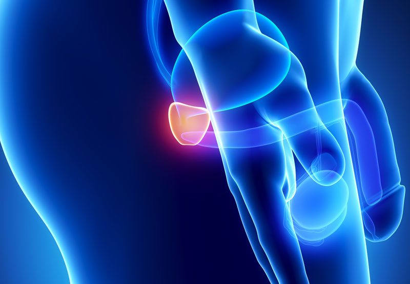

MRI-fusion targeted prostate biopsy is an advanced diagnostic procedure used to evaluate suspicious areas of the prostate for cancer. The technique combines previously obtained magnetic resonance imaging (MRI) with real-time ultrasound during the biopsy. By merging these images, the physician can precisely target lesions seen on MRI while also obtaining additional samples when appropriate. This method can improve detection of clinically significant prostate cancer compared with standard biopsy alone.
What It Treats
MRI-fusion targeted biopsy is used to diagnose or further evaluate:
- Elevated PSA levels
- Abnormal prostate MRI findings
- Abnormal digital rectal exam (DRE)
- Prior negative biopsy with continued concern for cancer
- Active surveillance monitoring for known low-risk prostate cancer
- Need for more accurate lesion targeting before treatment planning
How the Procedure Works
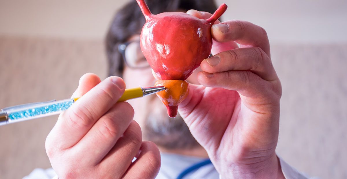
A prostate MRI is performed before the biopsy to identify suspicious regions. During the procedure, MRI images are electronically fused with live ultrasound images. This allows the physician to guide biopsy needles directly into the targeted area while also sampling standard zones of the prostate if needed.
Biopsies may be performed through:
- Transrectal approach: Through the rectal wall into the prostate
- Transperineal approach: Through the skin between the scrotum and rectum
Local anesthesia, sedation, or general anesthesia may be used depending on the biopsy method and patient needs.
Benefits of the Procedure
MRI-fusion targeted biopsy may offer several advantages:
- Improved accuracy when sampling suspicious MRI lesions
- Better detection of clinically significant prostate cancer
- Reduced sampling of insignificant areas in selected patients
- Helpful after a prior negative biopsy with persistent elevated PSA
- Useful for treatment planning and active surveillance monitoring
- Supports transperineal biopsy options with lower infection risk
What to Expect
Most biopsies are outpatient procedures. Temporary blood in the urine, stool, or semen is common for a short period after the procedure. Mild soreness or urinary symptoms may occur for several days. Antibiotics may be recommended depending on the biopsy route. Pathology results are reviewed during follow-up to discuss whether surveillance, repeat testing, or treatment is needed.
Is It Right for You?
MRI-fusion targeted prostate biopsy may be appropriate for men with elevated PSA, suspicious MRI findings, abnormal prostate exam results, prior negative biopsy, or those being monitored on active surveillance. The best biopsy route and technique depend on prostate anatomy, infection risk, imaging findings, and overall health. A urologist can determine whether this approach is the most effective next step.