

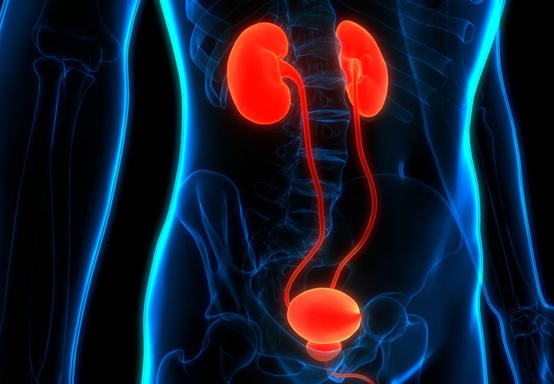
Upper tract urothelial carcinoma is a cancer that develops in the lining of the kidney drainage system (renal pelvis) or ureter, the tube that carries urine from the kidney to the bladder. It arises from the same urothelial cells found in the bladder, but occurs higher in the urinary tract. This type of cancer is less common than bladder cancer and may range from low-grade, slow-growing tumors to aggressive disease that can invade surrounding tissue or spread. Early diagnosis is important to preserve kidney function and improve treatment outcomes.
Causes
Several factors are associated with an increased risk of upper tract urothelial carcinoma, including:
- Smoking: Tobacco exposure is a major risk factor
- Prior bladder cancer: Patients with bladder tumors have a higher risk of related tumors elsewhere in the urinary tract
- Chemical exposure: Certain industrial chemicals may increase risk
- Chronic inflammation: Recurrent infections, stones, or long-term irritation may contribute
- Inherited syndromes: Lynch syndrome and other genetic conditions can raise risk
- Age: More common in older adults
- Personal history of urothelial cancer: Previous urinary tract tumors increase recurrence risk

Symptoms
Symptoms may be mild or intermittent. Common signs include:
- Blood in the urine (hematuria)
- Flank or side pain
- Recurrent urinary tract infections
- Kidney swelling from obstruction (hydronephrosis)
- Urinary urgency or frequency
- Unexplained weight loss or fatigue in advanced disease
Some tumors are found during evaluation for microscopic hematuria or abnormal imaging.
Diagnosis
Evaluation of upper tract urothelial carcinoma often includes:
- Urinalysis: Detects blood or infection
- Urine cytology: Looks for abnormal urothelial cells
- CT urogram: Common imaging study to evaluate kidneys, ureters, and bladder
- MRI: Used in selected patients when needed
- Cystoscopy: Examines the bladder because related tumors may occur there
- Ureteroscopy with biopsy: Direct visualization and tissue sampling of the tumor
- Staging imaging: Performed when invasive or advanced disease is suspected
Treatment Options
Treatment depends on tumor grade, size, location, kidney function, and overall health:
- Nephroureterectomy: Removal of the kidney, ureter, and bladder cuff for many higher-risk tumors
- Kidney-sparing endoscopic treatment: Laser ablation or resection for selected low-grade tumors
- Segmental ureterectomy: Removal of part of the ureter with reconstruction in selected patients
- Intraluminal therapy: Medication delivered into the urinary tract in selected cases
- Chemotherapy or immunotherapy: Used before surgery, after surgery, or for advanced disease
- Surveillance: Ongoing imaging and cystoscopy due to recurrence risk
When to See a Specialist
Patients with blood in the urine, unexplained hydronephrosis, flank pain, or imaging showing a ureter or renal pelvis abnormality should be evaluated promptly by a urologist. Early specialist care can help preserve kidney function, clarify diagnosis, and determine the most effective treatment plan.